 Atrijum Laboratory
 Physical Therapy
ECG
What is ECG? Electrocardiogram (ECG) represents a simple test used to check heart rhythm and cardiac activity. Signals sent by…


Ultrasound represents a non-invasive test, more precisely a medical diagnostic method. The ultrasound image alternatively called sonogram. Ultrasound uses high-frequency sound waves to create images or video recordings of internal organs or other soft tissues, such as blood vessels, in real time. Ultrasound enables healthcare workers to “see” soft tissue details within your body without making any incisions. Unlike X-rays, ultrasound uses no radiation. Although most people associate ultrasound with pregnancy, healthcare workers use ultrasound for various situations and to examine several different parts of your body’s interior.
Heart ultrasound represents a non-invasive procedure used to assess heart structure and function. During the procedure, the physician applies gel to the patient’s chest and uses an ultrasound probe emitting sound waves toward the heart. These waves reflect back to the probe, and computer program converts these reflections into heart images.
Heart ultrasound enables physicians to assess heart size, function and wall motility, heart chamber size, heart valve appearance and function, heart muscle shape and function, and presence of irregularities such as tumors or anomalies. This diagnostic method proves painless and safe, enabling physicians precise heart health assessment. Heart ultrasound can be used to diagnose various cardiac conditions such as heart failure, heart attack, arrhythmias, congenital cardiac anomalies, and other heart diseases.
Heart ultrasound, also known as echocardiography, typically performed on patients experiencing cardiac symptoms such as shortness of breath, chest pain, high blood pressure, or rapid heartbeat. During examination, patient lies on bed while technician moves sensor over chest. Sensor emits sound waves reflecting off heart, creating heart image on computer screen.
Heart ultrasound can detect wide range of cardiac diseases including:
In addition to diagnosing cardiac diseases, heart ultrasound can also help physicians monitor patient progress during cardiac disease therapy. This procedure frequently used for assessing heart condition during pregnancy.
Heart ultrasound with color Doppler, thanks to new technical capabilities, enables great amount of information about heart and cardiac structures such as: heart chamber dimensions, heart muscle dimensions, its motility, heart muscle strength to eject blood peripherally and relax, heart valve function, provides insight into pericardium (heart sac) condition.
Heart ultrasound using color Doppler method enables insight into blood flow velocities through cardiac orifices and diagnosis of congenital or acquired heart defects. Physician, adequately trained for this diagnostic type, moves ultrasound device probe over skin of left chest side, during which patient feels no pain. Examination lasts 20 to 30 minutes, after which patient receives explanation and written report. Emphasis proves that expert heart ultrasound provides much more information than basic.
Expert heart ultrasound performed using concrete calculation and quantification method of heart muscle motility in three directions, with only one heart quarter (left ventricle) examined in 17 segments, each separately. This proves particularly important examination providing additional and fuller information for patients who had heart attack, have angina pectoris, elevated blood pressure, and heart failure.
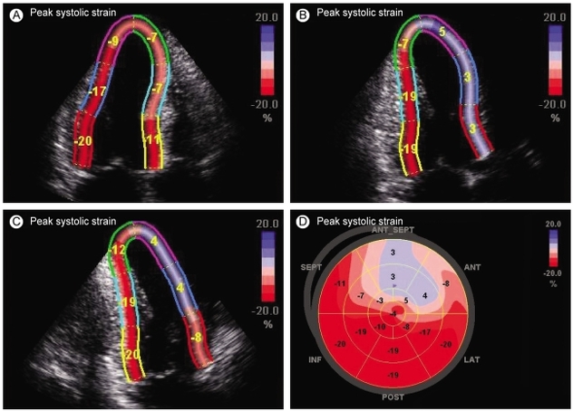
Thus assessed reduced heart muscle motility may indicate high likelihood of occlusion or narrowing of specific artery supplying heart muscle.
Until now, such heart blood vessel examination performed only by coronary angiography; now we have ultrasound examination able to provide adequate data and refer patient for potential therapeutic coronary angiography involving stent placement.
Large number of patients can avoid coronary angiography (catheterization) using this method, concerning patients needing such procedure for diagnosis.
Additionally, extremely important that expert heart ultrasound, just like basic ultrasound examination, proves safe for patient, painless, without harmful radiation and contrast agents, requiring no special preparation.
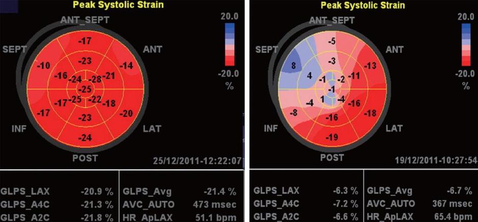
Expert method heart ultrasound provides currently most complete heart picture, not requiring invasive approach exposing patient to radiation, contrast agents, hospital stay, and special examination preparation.
Also worth mentioning how such examination represents standard for patients after stent or bypass implantation, for assessing aortic stenosis and its further monitoring, and for oncology patients, heart failure patients, and myocardial hypertrophy (thickening) from either sports or pathological causes.

Recommend this service

What is ECG? Electrocardiogram (ECG) represents a simple test used to check heart rhythm and cardiac activity. Signals sent by…
What is ECG? Electrocardiogram (ECG) represents a simple test used to check heart rhythm and cardiac activity. Signals sent by…